

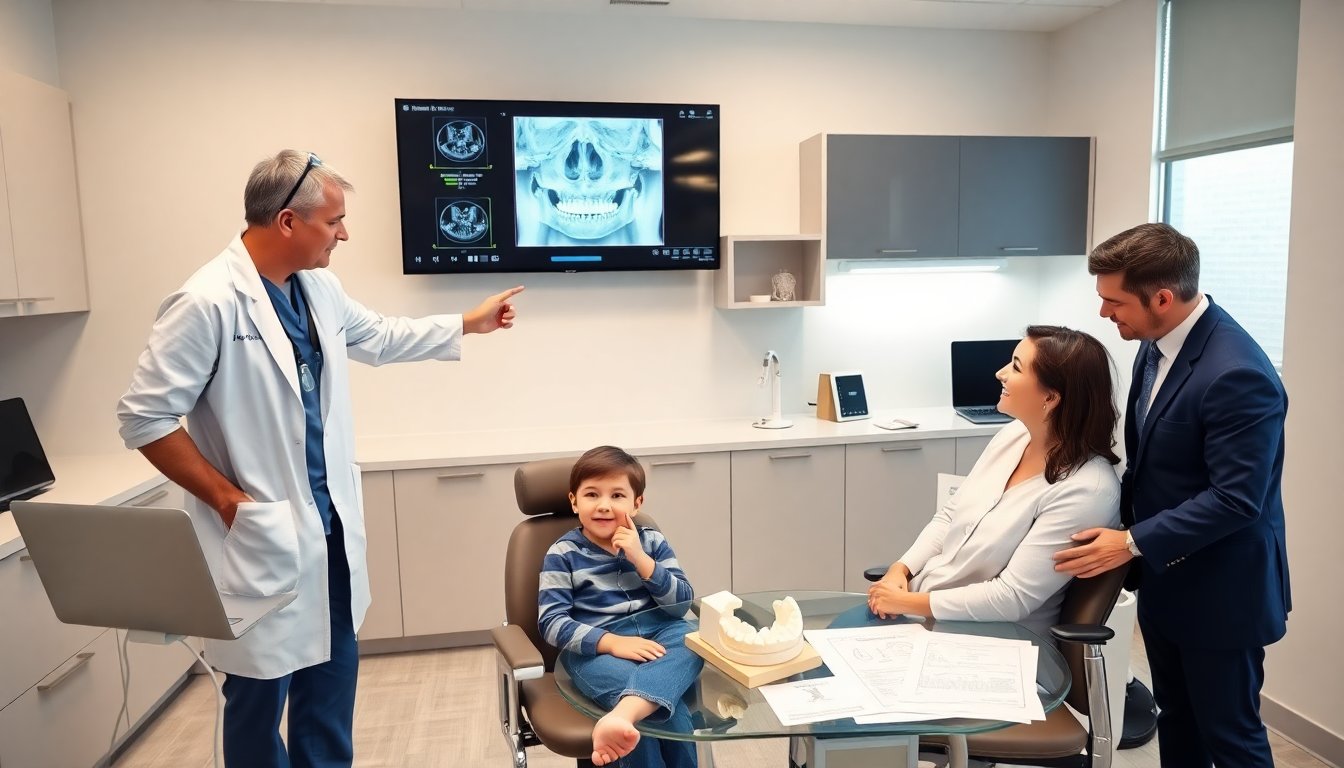
Syndrathia appears early in life and it causes parts of the jaw to fuse. This condition limits mouth opening and affects eating and speech. Clinicians need clear criteria to diagnose syndrathia. Families often seek surgical care and long-term therapy. This guide covers definition, clinical signs, causes, imaging, surgery, and recovery. It gives concise, actionable information for clinicians, parents, and allied professionals.
Key Takeaways
- Syndrathia is a rare condition involving fusion of the jaws that limits mouth opening and impacts feeding and speech.
- Early diagnosis using clinical evaluation and imaging like CT scans is critical for effective management of syndrathia.
- Treatment includes conservative therapy for fibrous fusion and surgical release for bony or dense fibrous fusion to restore function.
- Surgical timing depends on patient’s age, fusion type, and airway risk, with techniques like gap arthroplasty and coronoidectomy commonly used.
- Multidisciplinary care involving dentistry, nutrition, speech therapy, and psychology improves outcomes and supports families.
- Postoperative rehabilitation focusing on jaw exercises and speech therapy is essential to prevent refusion and enhance functional recovery.
What Is Syndrathia? Definition, Types, And Epidemiology
Syndrathia refers to partial or complete fusion between the mandible and maxilla or between the jaws and the cranial base. It occurs in congenital or acquired forms. Congenital syndrathia often appears with other craniofacial anomalies. Acquired syndrathia follows trauma, infection, or prolonged immobilization. Clinicians classify syndrathia as fibrous, bony, or mixed fusion. Fibrous fusion shows connective tissue between bones. Bony fusion shows osseous bridging. Mixed fusion has both elements. Incidence is low. Case reports and small series provide most data. The condition appears slightly more often in males in published series.
Clinical Presentation And Diagnosis
Patients with syndrathia present with limited mouth opening, feeding difficulty, and speech delay. Infants may show poor weight gain. Children may have facial asymmetry and dental malocclusion. Clinicians perform history and focused oral exam. They note range of motion, presence of palpable bony ridges, and oral hygiene. They document airway status and nutritional intake. Early recognition helps plan treatment. Referral to a craniofacial or maxillofacial team improves outcomes. Families need clear counseling about the condition and the diagnostic steps.
Diagnostic Imaging And Clinical Assessment
CT imaging gives high-resolution detail of bone fusion in syndrathia. Cone beam CT reduces radiation and shows jaw anatomy well. MRI helps when the clinician suspects soft tissue or fibrous bands. Ultrasound can assess superficial soft tissue in infants. Clinicians order dental models and occlusal analysis to plan surgery. Video fluoroscopy can evaluate swallow when feeding is impaired. The team combines imaging with functional measures to decide timing and approach. Preoperative planning often uses 3D models and virtual surgical planning.
Causes, Genetics, And Associated Anomalies
Congenital syndrathia may relate to genetic syndromes or in utero events. Mutations in craniofacial development genes appear in some reports. Clinicians test for syndromic features such as ear anomalies, limb defects, or cardiac issues. Acquired causes include facial trauma, postoperative scarring, infection, and prolonged ankylosis after immobilization. Syndrathia frequently associates with micrognathia, cleft palate, and ear malformations. Genetic counseling helps families when syndromic signs appear. The team screens the child for other anomalies to guide treatment and prognosis.
Treatment Strategies And Surgical Options
Treatment for syndrathia aims to restore mouth opening, protect the airway, and support growth. Clinicians start with conservative care for fibrous bands when possible. Conservative care includes physical therapy and jaw stretching devices. Surgical release becomes necessary for dense fibrous or bony fusion. Surgeons plan procedures to avoid damage to teeth, nerves, and growth centers. The team may stage interventions to balance function and facial growth. Multidisciplinary care includes dentistry, nutrition, speech therapy, and psychology to support the child and family through treatment.
Surgical Timing And Common Operative Techniques
Surgeons decide timing based on age, fusion type, and airway risk. They delay major reconstruction when growth allows unless the airway or nutrition is compromised. Common techniques include gap arthroplasty, interpositional grafting, and coronoidectomy. Surgeons use autologous tissue or alloplastic material to prevent refusion. Endoscopic-assisted release can reduce morbidity in select cases. Virtual surgical planning and intraoperative navigation improve precision. Surgeons plan for possible staged bone grafting or distraction osteogenesis when the defect threatens facial form.
Postoperative Care, Rehabilitation, And Prognosis
After surgery, clinicians monitor airway and pain closely. Early range-of-motion exercises reduce the risk of refusion in syndrathia. Physical therapists lead jaw stretching and guided exercises. Speech therapists address articulation and swallowing. Dentists manage occlusion and dental development. The team schedules serial imaging and clinic visits to track growth and detect recurrence. Prognosis depends on fusion type, associated anomalies, and timing of intervention. Many patients gain significant function with coordinated care, but some require further procedures during growth.